Tiedosto:Hepatocellular carcinoma 1.jpg
Hepatocellular_carcinoma_1.jpg (550 × 368 kuvapistettä, 38 KiB, MIME-tyyppi: image/jpeg)
| Tämä tiedosto on tiedostotietokanta Wikimedia Commonsista. Tiedot kuvaussivulta näkyvät alla. |  |
Tiedoston kuvaussivu Commonsissa |
Yhteenveto
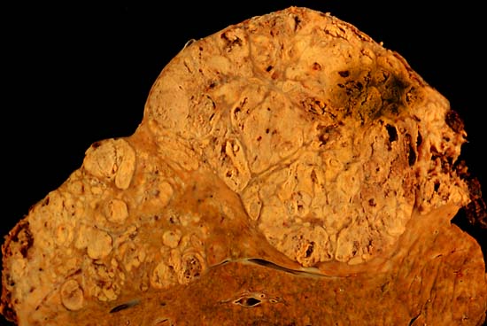
| Kuvaus |
Hepatocellular carcinoma This specimen is from a 50ish woman who presented to the hospital with abdominal pain and ascites. The radiologist recovered what appeared to be whole blood on paracentesis. Cytological exam of the bloody fluid showed no evidence of malignancy. Liver function tests were abnormal, and serologic tests were positive for antibody to hepatitis C. The patient deteriorated rapidly and died within a few days. The autopsy showed this hepatocellular carcinoma occupying much of the volume of a cirrhotic liver. Furthermore, the tumor had invaded the diaphragm and ruptured into the peritoneal cavity, causing the bloody ascites. The photo shows a view of a longitudinal slice taken through the full length of the liver. The photos were shot with a Minolta X-370 with 100 mm bellows lens on Kodak Elite ISO 100 transparency film. The specimen was sliced fresh and fixed in formalin overnight, then briefly immersed in 70% alcohol to retrieve some of the native color and dull the surface reflections. Photograph by Ed Uthman, MD. Public domain. Posted 23 Sep 00 |
| Lähde | http://web2.airmail.net/uthman/specimens/index.html |
| Tekijä | |
| Käyttöoikeus (Tämän tiedoston uudelleenkäyttö) |
PD |
Lisenssi
| Tämän teoksen tekijä, Ed Uthman, on julkaissut sen public domainiin. Tämä on voimassa maailmanlaajuisesti. Joissain maissa laki ei mahdollista tätä. Mikäli näin on: Ed Uthman myöntää kaikille oikeuden käyttää tätä teosta mihin tahansa tarkoitukseen ilman minkäänlaisia ehtoja, ellei laki vaadi ehtojen asettamista.
|
Tiedoston historia
Päiväystä napsauttamalla näet, millainen tiedosto oli kyseisellä hetkellä.
| Päiväys | Pienoiskuva | Koko | Käyttäjä | Kommentti | |
|---|---|---|---|---|---|
| nykyinen | 5. kesäkuuta 2006 kello 13.14 | | 550 × 368 (38 KiB) | Patho | {{Information| |Description=Hepatocellular carcinoma This specimen is from a 50ish woman who presented to the hospital with abdominal pain and ascites. The radiologist recovered what appeared to be whole blood on paracentesis. Cytological exam of the blo |
Tiedoston käyttö
Seuraavat 2 sivua käyttävät tätä tiedostoa:
Tiedoston järjestelmänlaajuinen käyttö
Seuraavat muut wikit käyttävät tätä tiedostoa:
- Käyttö kohteessa ar.wikipedia.org
- Käyttö kohteessa ast.wikipedia.org
- Käyttö kohteessa az.wikipedia.org
- Käyttö kohteessa be.wikipedia.org
- Käyttö kohteessa bs.wikipedia.org
- Käyttö kohteessa ca.wikipedia.org
- Käyttö kohteessa cs.wikipedia.org
- Käyttö kohteessa de.wikipedia.org
- Käyttö kohteessa de.wikibooks.org
- Käyttö kohteessa el.wikipedia.org
- Käyttö kohteessa en.wikipedia.org
- Hepatocellular carcinoma
- Alcohol and cancer
- Portal:Medicine/Selected article/50, 2007
- Portal:Medicine/Selected Article Archive (2007)
- Obesity-associated morbidity
- Cirrhosis
- Portal:Viruses
- Portal:Viruses/Selected article
- Portal:Viruses/Selected article/10
- User:Daniel Mietchen/Wikidata lists/Items with Disease Ontology IDs
- Käyttö kohteessa eo.wikipedia.org
- Käyttö kohteessa es.wikipedia.org
- Käyttö kohteessa eu.wikipedia.org
- Käyttö kohteessa fa.wikipedia.org
- Käyttö kohteessa fr.wikipedia.org
- Käyttö kohteessa gl.wikipedia.org
- Käyttö kohteessa he.wikipedia.org
- Käyttö kohteessa hi.wikipedia.org
- Käyttö kohteessa hy.wikipedia.org
- Käyttö kohteessa id.wikipedia.org
- Käyttö kohteessa it.wikipedia.org
- Käyttö kohteessa ja.wikipedia.org
- Käyttö kohteessa kk.wikipedia.org
Näytä lisää tämän tiedoston järjestelmänlaajuista käyttöä.